
FIG 4.
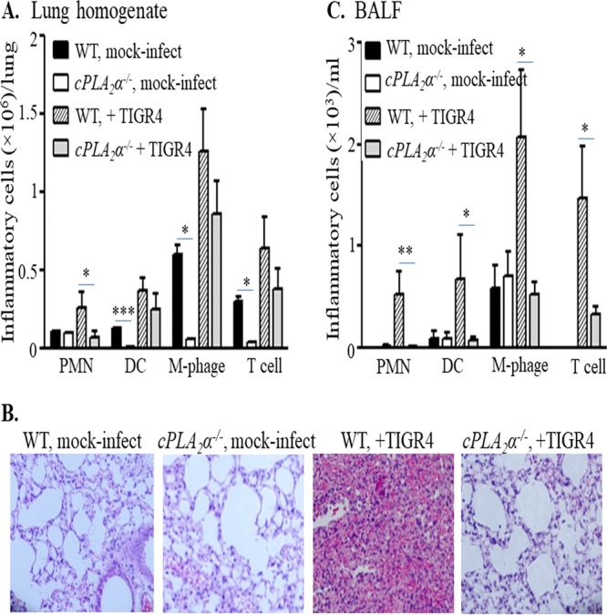
cPLA2α promotes pulmonary inflammation in S. pneumoniae-infected mice. Mice (cPLA2α−/− or their WT littermates) were mock infected with PBS (n = 4 for each strain, per experiment) or infected intratracheally with S. pneumoniae TIGR4 (n = 5 for each strain, per experiment), as detailed in Materials and Methods. Lungs and bronchoalveolar lavage fluid (BALF) were collected from the mock-infected or TIGR4-infected mice. Cells present in the digested lungs and BALF were stained with relevant MAbs, and the fluorescence intensities of the stained cells were determined by flow cytometry. Collected data were analyzed to determine the numbers of PMNs, dendritic cells (DC), macrophages (M-phage), or T cells in the lungs (A) and in the BALF (C). Statistical significance was analyzed by one-way ANOVA followed by individual Student's t test analyses. *, P < 0.05; **, P < 0.005; ***, P < 0.0005. For histological analyses (B), H&E-stained lung sections were prepared and examined by light microscopy (original magnification, ×20). Shown are results from a representative of two independent experiments.