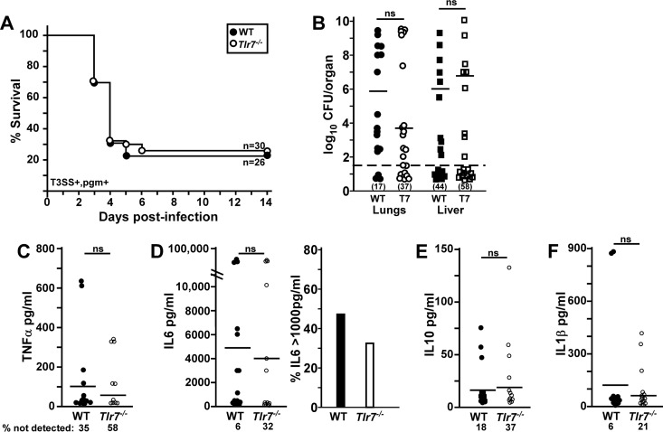
FIG 6.
Tlr7−/− mice experience reduced serum TNF-α levels and sepsis during Y. pestis CO92 infection. Groups of 10 WT and Tlr7−/− mice were challenged by intranasal infection with 1 × 10³ CFU Y. pestis CO92 (T3SS+ pgm+). (A) Survival was monitored over 14 days. Data shown were collected in 3 independent trials (n = 26 or 30 per group). (B to F) On day 3 postinfection, animals were euthanized; blood was collected by cardiac puncture for multiplex serum cytokine analysis; and lungs, liver, and spleen were homogenized for the enumeration of the bacterial load. (B) Bacterial counts in the indicated tissues. Bars indicate medians, and the percentages of each tissue that had undetectable bacterial titers are indicated in parentheses. (C) Serum TNF-α. (D) IL-6. The right panel shows percentages of animals in each group with high IL-6 titers. (E) IL-10. (F) IL-1β. Bars indicate the means, and the percentages of mice with undetectable cytokines are indicated (n = 17 WT and 19 Tlr7−/− mice [3 WT mice and 1 Tlr7−/− mouse died prior to 72 hpi], collected in two independent trials). Statistical significance was evaluated by a Mantel-Cox log rank test (A) or by Student's t test (B to F). ns, not significant.

