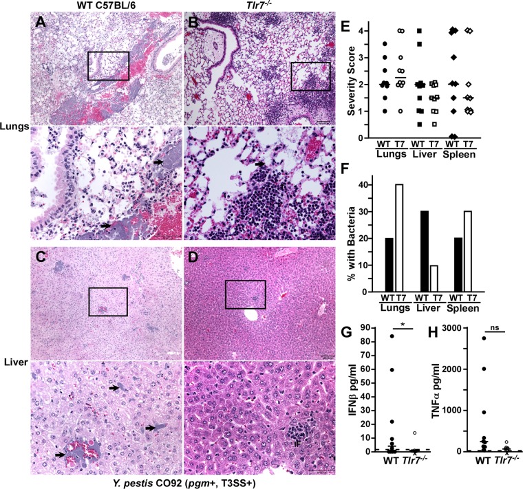
FIG 7.
TLR7 is associated with increased liver pathology during pneumonic plague. (A to F) Groups of 5 WT (A, C, E, and F) or Tlr7−/− (B, D, E, and F) mice were challenged by intranasal infection with 1 × 10³ CFU Y. pestis CO92 (T3SS+ pgm+). (A to D) On day 3 postinfection, animals were euthanized, and formalin-fixed lungs (A and B), liver (C and D), and spleen (see Fig. S4A and S4B in the supplemental material) were sectioned, stained with hematoxylin and eosin, and analyzed by histopathology. Representative lesions are shown; boxes outline the zoomed-in areas shown in the bottom panels, and arrows point to bacteria in the zoomed-in images. Bars, 100 μm (50 μm in the zoomed-in images). (E and F) Quantification of histopathology. (E) Severity scoring; (F) percentages of samples with bacterial microcolonies visible by histopathology. Bars indicate medians. (G and H) Serum IFN-β and TNF-α levels were measured by an ELISA. Bars indicate medians, dotted lines indicate the limit of detection, and X indicates that the animal died prior to analysis. Statistical significance was evaluated by Student's t test. *, P < 0.05. Data shown in all panels were collected in 2 independent trials (n = 10 per group).

