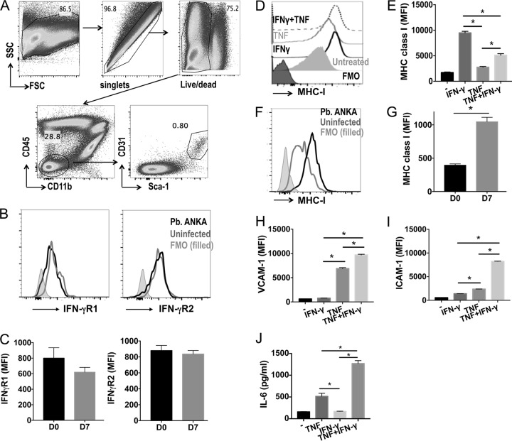
FIG 6.
IFN-γ promotes direct activation of brain endothelial cells under inflammatory conditions. WT mice were infected i.v. with 104 P. berghei ANKA pRBCs. Brains were removed from infected mice on day 7 of infection and from naive mice. (A to C) Representative plots showing gating strategy to identify brain endothelial cells (A), representative histograms (B), and mean fluorescence intensities (MFI) of IFN-γR1 and IFN-γR2 expression by brain endothelial cells from infected and naive mice (C). FSC, forward scatter; SSC, side scatter. (D, E) Brain endothelial cells (bEnd5 cell line) were cultured in vitro and activated for 18 h with TNF (1 ng/ml) and/or IFN-γ (1 ng/ml). Representative flow cytometry histogram (D) and calculated mean MFI (E) of MHC class I expression by unstimulated and stimulated bEnd5 cells. (F, G) Representative histogram (F) and MFI (G) showing MHC class I expression by brain endothelial cells from P. berghei ANKA-infected and naive mice. (H to J) MFI of VCAM-1 (H) and ICAM-1 (I) on and production of IL-6 (J) by stimulated and unstimulated bEnd5 cells, measured by ELISA. Results in panels C and G are the mean values ± SEM of the groups, with 5 mice per group. Results in panels E and H to J are the mean values ± SEM from three independent biological replicates and are representative of 2 independent experiments. *, P < 0.05 between defined groups. Statistical significance was tested using the Mann-Whitney test for the data in panels C and G and one-way ANOVA with Tukey's post hoc test for the data in panels E and H to J.

