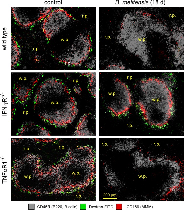
FIG 7.
Visualization of marginal zone macrophage populations in control and infected wild-type and deficient mice. Wild-type, TNF-αR1−/−, and IFN-γR−/− C57BL/6 mice were infected i.p. with a dose of 105 CFU of B. melitensis and sacrificed at the indicated time postinfection. The panels show the localization of CD45R/B220+ B cells, CD169+ cells (MMMs), and FITC-coupled dextran+ cells (MZMs) on spleen sections from PBS-treated mice (control) and mice at 18 days postinfection. The panels are color coded with the text for the antigen examined. r.p., red pulp; w.p., white pulp. These results are representative of at least two independent experiments (n = 6).

