
Fig. 1.
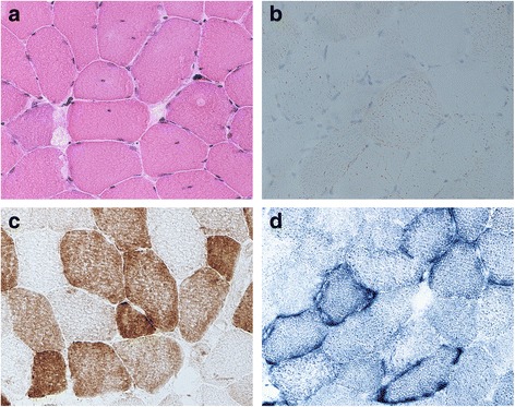
Muscle histology and histochemistry. a H&E stain revealing an enhanced variability in fiber size and some fibers showing internalized nuclei or increased subsarcolemmal eosinophilic accumulation. b Oil-Red-O stain revealing increased amount of lipid droplets. c COX histochemistry demonstrating a significant number of COX-deficient fibers. d SDH histochemistry showing multiple ragged-blue-fibers with subsarcolemmal accumulation of abnormal mitochondria