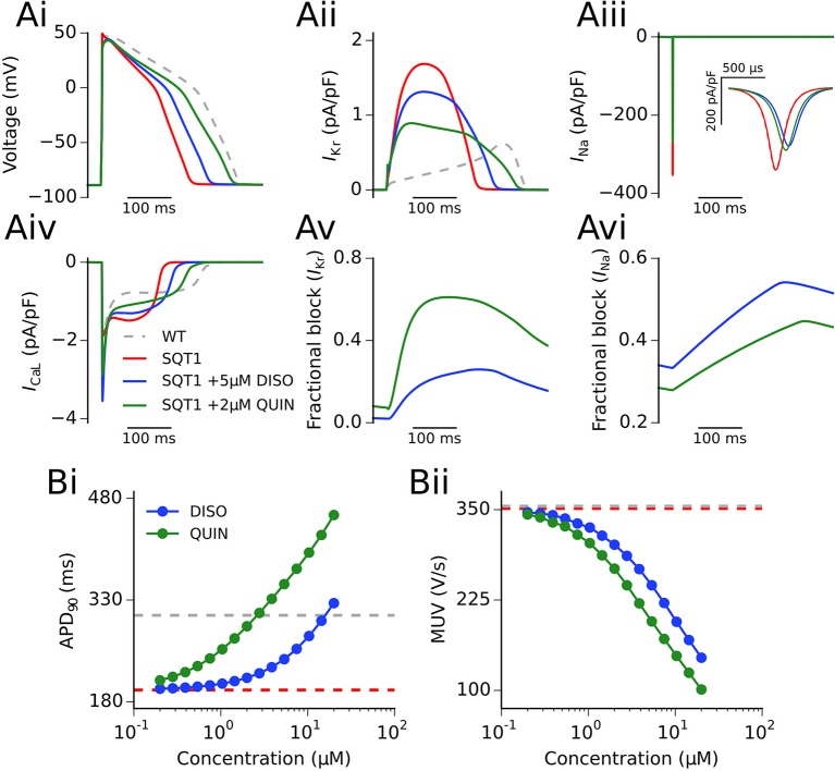
Figure 3.
Single cell action potentials and current traces. (Ai) Endocardial action potential (AP) at 1 Hz under WT (silver, dashed line), SQT1 (red, solid line), SQT1 + 5μM disopyramide (DISO) (blue, solid line), and SQT1 + 2μM quinidine (QUIN) (green, solid line) conditions. Corresponding current traces are shown for IKr (Aii), INa (Aiii), and ICaL (Aiv). The degree of fractional block of IKr and INa is shown in (Av,Avi), respectively. The concentration dependence of the single cell AP duration at 90% repolarization (APD90) and maximum upstroke velocity (MUV) is shown in (Bi,Bii), respectively.

