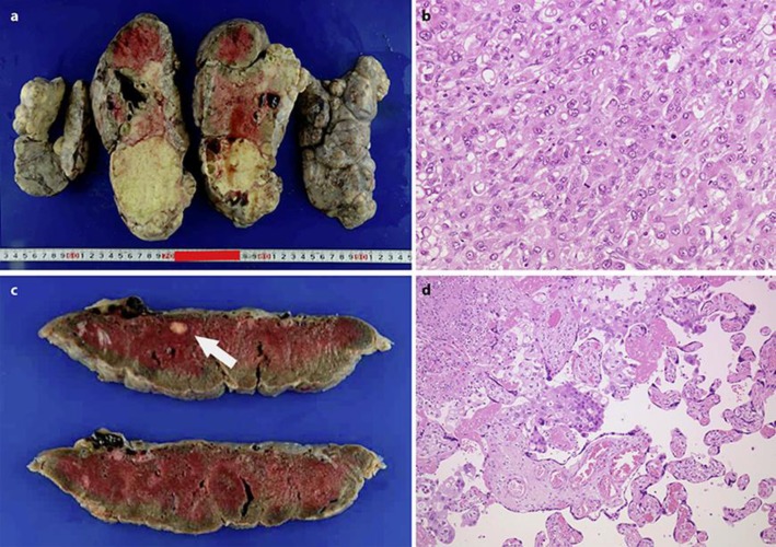
Fig. 3.
a Macroscopic findings of the left ovary resected at our hospital. b Pathological findings of the left ovary resected at our hospital. The left ovarian tumour was composed of poorly differentiated adenocarcinoma, the characteristics of which were identical to those observed in the previously resected right ovarian tumour (magnification ×20). c Macroscopic findings of the placenta. A white nodule measuring 1 × 1 cm was observed in the specimen. d Pathological findings of placenta. Characteristics and appearance of cancer cells observed in the placenta were similar to those observed in the ovarian tumours (magnification ×40).

