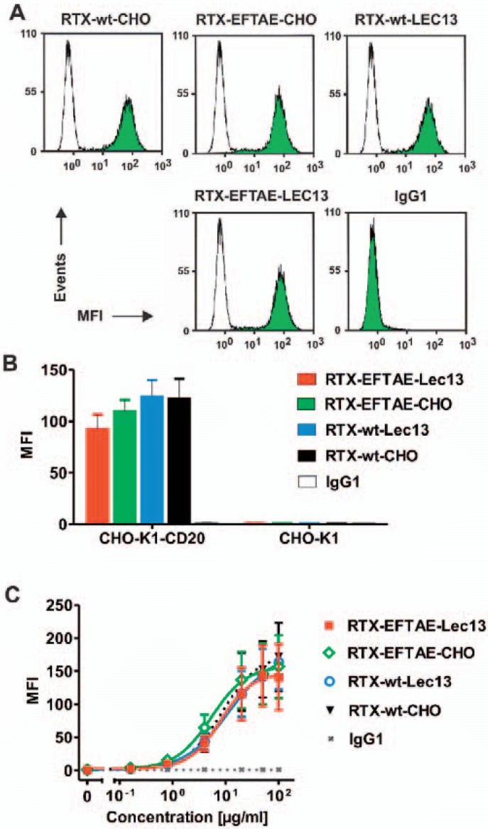
Fig. 2.
CD20 binding analysis. A CD20-positive MEC-2 cells were incubated in buffer alone (white peaks) or in the presence of the indicated antibodies at 50 µg/ml (green peaks), then reacted with FITC-conjugated anti-human IgG Fc F(ab')2 and analyzed by flow cytometry. B RTX-wt-CHO, RTX-EFTAE-CHO, RTX-wt-Lec13 and RTX-EFTAE-Lec13 (concentration: 50 µg/ml) specifically bound to CHO-K1-CD20 cells but did not react with non-transfected CHO-K1 cells. Bars indicate mean values ± SEM (n = 2). Antibodies were detected with FITC-conjugated anti-human IgG Fc F(ab')2 fragments and flow cytometry. Trastuzumab was used as control antibody (MFI, mean fluorescence intensity). C Antibody variants were analyzed for binding to CHO-K1-CD20 cells at varying concentrations using secondary FITC-conjugated anti-human IgG Fc F(ab')2 fragments for detection and flow cytometry. Data points represent mean values ± SEM (n = 4).

