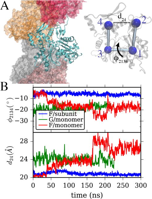
Figure 1.

(A) Left, a snapshot from an actin filament simulation shows one actin subunit in ribbon style with a bound ATP molecule, surrounded by adjacent subunits. Right, a single actin subunit is overlaid with CG beads at the center of mass of its four major subdomains. Important CVs describing the transition from globular to filamentous conformation are the “cleft distance” from bead 2 to 4, and the “twist” dihedral angle formed by the four subdomains, a rotation around the central “bond” as shown. (B) The values of twist angle and cleft distance are shown for three systems. In blue is a single actin subunit within a filament, in green, a single G-actin in solution starting from its crystal structure, and in red, a single actin in solution starting from the filamentous structure.
