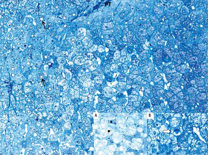
Fig. 1.
Light microscopic observations of semithin epon sections (toluidine blue stain; original magnification 200×). Mild hydropic degeneration of hepatocytes of periportal zone. The hepatocytes have pale-staining, rare cytoplasm. Portal tract (PT), “empty” nucleus (black arrow), lipid droplet (white arrow). A) Two basic types of hepatocellular changes: hydropic degeneration (•) and acidophilic degeneration (♦). B) Hepatocyte mitosis (white bold arrow)

