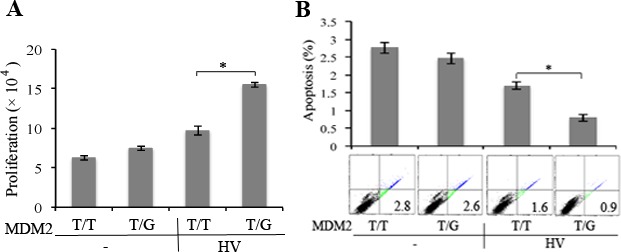
Figure 2.

HV enhanced proliferation and survival of hRPE cells with MDM2 T309G compared with those with MDM2 T309T only. (A) The hRPE cells with MDM2 T309 only (T/T) or MDM2 T309 plus G309 (T/G) were plated into a 24-well plate at a density of 3 × 10⁴ cells per well. After the cells had attached the plates, the medium was switched to either DMEM/F12 or HV (diluted 1:3 in DMEM/F12). The media were replaced every day. On day 3, cells were counted with a hemocytometer under a light microscope. Mean ± SD of three independent experiments is shown. *P < 0.05, unpaired t-test. (B) Serum-starved hRPE cells with MDM2 T309G or T309T only were plated into 60-mm dishes at a density of 100,000 cells per dish. After the cells attached to the dishes, the medium was switched to either DMEM/F12 or HV (diluted to 1:3 in DMEM/F12). The media were replaced every day. On day 3, the cells were stained with FITC-conjugated annexin V and PI in an apoptosis assay kit by following the manufacturer's instructions. Cells that were stained with annexin V and/or PI were detected and quantified by flow cytometry in a Beckman Coulter (Brea, CA, USA) XL instrument. The mean ± SD of three independent experiments is shown, and one of the experimental raw data is shown below the bar graphs.
