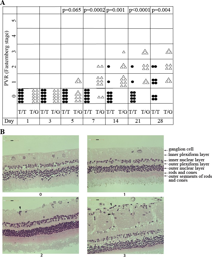
Figure 4.

The hRPE cells with MDM2 T309G had more potential to induce PVR in a rabbit model. (A) PVR was induced in the right eyes of Dutch Belted rabbits (10 rabbits per group) as previously described.25,27,28 Briefly, 1 week after gas vitrectomy, rabbits were intravitreally injected with 0.1 ml PRP and hRPE cells (3 × 10⁵) with MDM2 T309 only (T/T) or MDM2 T309 plus G309 (T/G) in 0.1 mL DMEM/F12. The rabbits were examined with an indirect ophthalmoscope and the PVR status for each rabbit was plotted on days 1, 3, 5, 7, 14, 21, and 28. The data were subjected to Mann-Whitney analysis. A P value less than 0.05 is considered a significant difference between the two groups (T/T and T/G). (B) The representative eyeballs from rabbits with PVR stages 0 and 1 (hRPE T309T), and 2 and 3 (hRPE T309G) were fixed, sectioned and stained with hematoxylin-eosin. In the eyes with PVR stages 2 and 3 injected with hRPE expressing MDM2 T309G, there were some cells (arrowheads) attaching to the retina.
