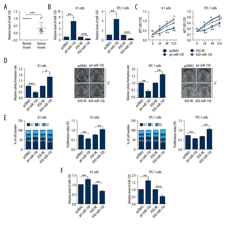
Figure 1.
MiR-150 was downregulated in thyroid cancer (TC) tissues and inhibited TC cell proliferation. (A) The expression level of miR-150 in thyroid tissues was determined by RT-qPCR assays. (B) The efficiency of pri-miR-150 expression plasmid or ASO-miR-150 was confirmed by RT-qPCR assays in TC cells. (C) The impacts of miR-150 on K1 and TPC-1 cellular viabilities were determined by MTT assays. (D) The relative colony formation rate of K1 and TPC-1 cells with indicated treatment was determined by colony formation assays. (E) The impacts of miR-150 on cell cycle of K1 and TPC-1 cells were evaluated by flow cytometry cell cycle assays. (F) The impacts of miR-150 on cell apoptosis of K1 and TPC-1 cells were evaluated by flow cytometry cell apoptosis assays; * p<0.05; ** p<0.01; *** p<0.001.

