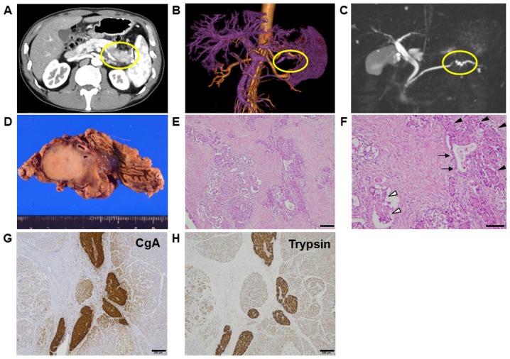
Figure 1.
(A) Abdominal computed tomography revealing a tumor 3 cm in diameter in the body of the pancreas. (B) Tumor enhancement from the splenic artery was indicated by magnetic resonance angiography. (C) Stenosis of the pancreatic duct was observed by magnetic resonance cholangiopancreatography. (D) Gross findings showed a round, hard tumor with a serrated margin in the body of the pancreas. The tumor measured 3 cm in diameter and was growing within the pancreatic parenchyma. (E-H) Pathologic examination revealed a mixed pancreatic carcinoma. Representative image of haematoxylin and eosin staining with (E) low-power and (F) high-power magnification. Cellular morphology indicated tumor components of ductal carcinoma (arrow), acinar cell carcinomas (ACC; black arrow head), and neuroendocrine carcinomas (NEC; white arrow head) in (F). Immunohistochemical analysis showed positive staining for chromogranin A (CgA) as a marker of (G) NEC and trypsin as a marker of (H) ACC. Scale bars, 200 µm for (E, G and H); 100 µm for (F).

