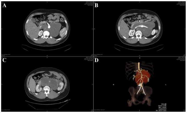
Figure 1.
CT angiogram images of the tumor demonstrate (A) encasement of the origin of the superior mesenteric artery, (B) encasement of the origins of the bilateral renal arteries and (C) involvement of the inferior vena cava at the level of right renal vein. (D) Reconstructed CT angiogram image of the tumor showing encasement of the origin of superior mesenteric artery and bilateral renal arteries. CT, computed tomography.

