
Figure 1. PHS decreases EMT in breast cancer cells.
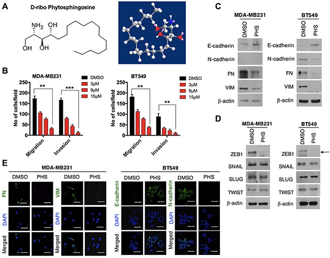
(A) Chemical and molecular structure of phytosphingosine (PHS) generated by ChemDraw professional software. (B) Invasion and migration assays of PHS-treated MDA-MB231 and BT549 basal-type breast cancer cells at various concentrations. (C) Western blot analysis of the EMT markers Fibronectin (FN), Vimentin (VIM), E-cadherin and N-cadherin in MDA-MB231 or BT549 cells after a DMSO or PHS treatment (10μM). (D) Western blot analysis of EMT transcription factors of SNAIL, SLUG, ZEB1 and TWIST in PHS (10μM)-treated MDA-MB231 and BT549 cells. (E) Immunofluorescence staining of EMT surface markers in PHS-treated MDA-MB231 and BT549 cells at a concentration of 10μM. DAPI, 4,6-diamidino-2-phenylindole. DMSO is used as a vehicle control here at a concentration similar to that of PHS. β-actin was used as a loading control. Scale bar = (100μm). Error bars represent the mean ± S.D. of triplicate samples. *p < 0.05, **p < 0.01 and ***p < 0.001.