
Figure 2. PHS suppresses the self-renewal ability of breast-like stem cell-populations.
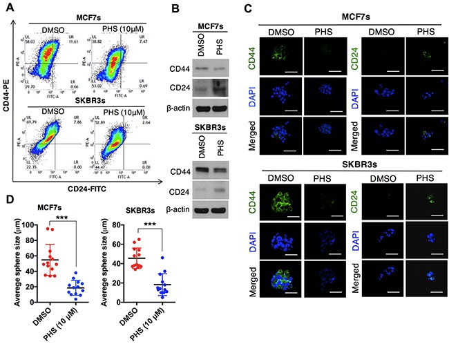
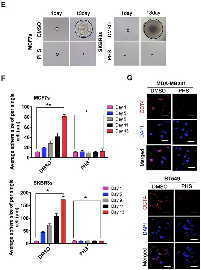
(A) FACS analysis for the CD44-PE and CD24-FITC expression levels in DMSO or PHS (10μM)-treated MCF7 (upper panel) and SKBR3 (lower panel) mammospheres. (B) Western blot analysis results of CD44 and CD24 protein levels in DMSO or PHS (10μM)-treated MCF7 and SKBR3 mammospheres. β-actin was used as a loading control. (C) Immunofluorescence staining of CD44 and CD24 expression levels in DMSO or PHS (10μM)-treated MCF7and SKBR3 mammospheres. (D) Quantification of the sphere-forming capabilities of MCF7 and SKBR3 sphere cells after a treatment with PHS (10μM) or a control vehicle, DMSO. (E) Clonal assay results of MCF7 and SKBR3 sphere cells 13 days after a treatment with PHS (10μM) or the control vehicle DMSO. (F) Changes in the sphere size of single cell in the presence of DMSO and PHS were monitored at different time points in both cells and are plotted on a graph. (G) Immunofluorescence staining of the OCT4 expression levels in DMSO or PHS (10μM)-treated MCF7 and SKBR3 spheres. Scale bar = (100μm). Error bars represent the mean ± S.D. of triplicate samples. *p < 0.05, **p < 0.01 and ***p < 0.001.