
Figure 2.
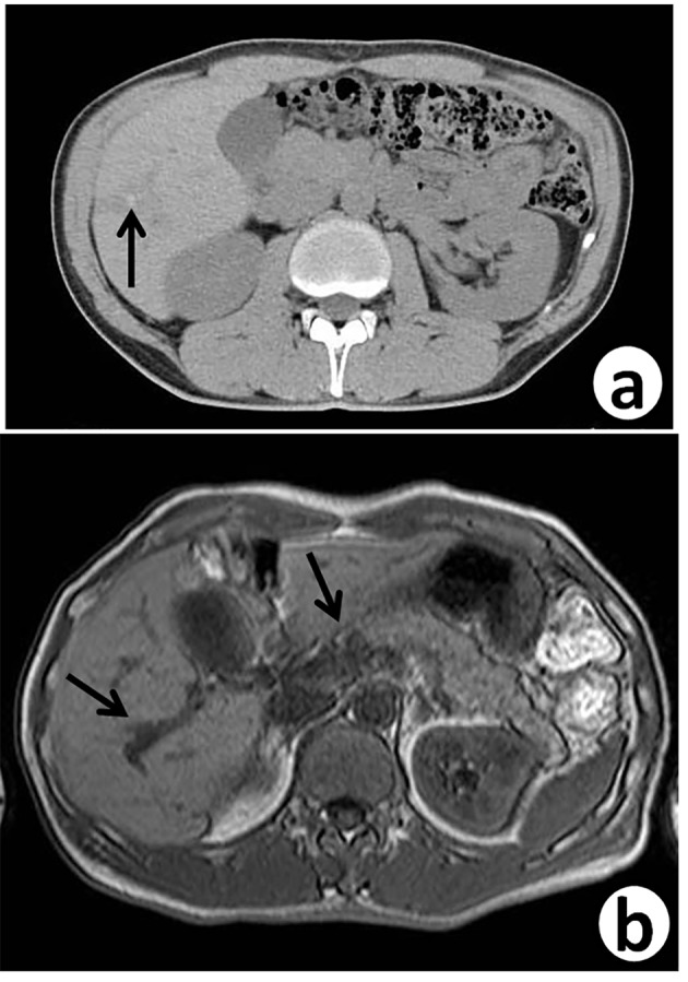
Follow-up abdominal contrast-enhanced CT scan (a) and MRI-T2 (b), 7 years after sigmoidectomy, reveal small dilated intrahepatic bile ducts (arrows) with thickening in the S5/6 area.
Official websites use .gov
A
.gov website belongs to an official
government organization in the United States.
Secure .gov websites use HTTPS
A lock (
) or https:// means you've safely
connected to the .gov website. Share sensitive
information only on official, secure websites.
Follow-up abdominal contrast-enhanced CT scan (a) and MRI-T2 (b), 7 years after sigmoidectomy, reveal small dilated intrahepatic bile ducts (arrows) with thickening in the S5/6 area.