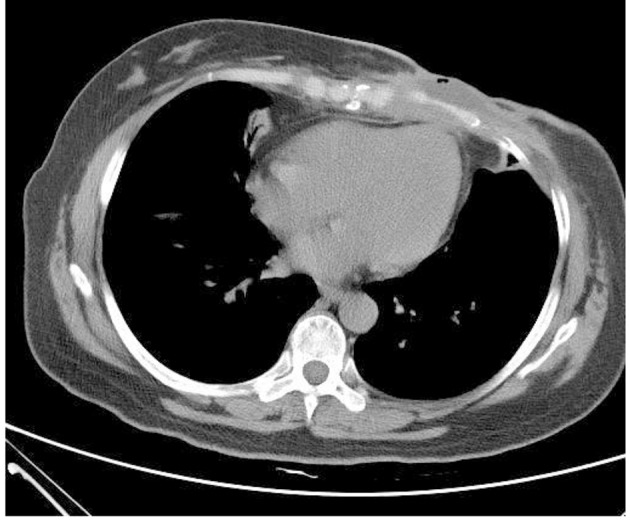
Figure 3.

Chest CT (on December 19, 2013): irregular low-density lesions were seen in front of sternum with the maximum cross-section of 4.3 × 2.9 cm. The border with the incrassated skin and the chest wall were not clear and the bone destruction was found in the sternum and the left fifth rib with the vague edge seen in part of costal cartilages.
