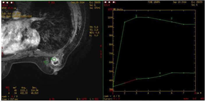
Figure 5.
Breast MRI (on September 19, 2014): one about 2.3 × 1.8 × 2.8 cm abnormal signal image with the irregular form was observed in the outer quadrant of the right breast with the equal T1 and long T2 signals. The border was unclear with the burr at the margin. The near skin incrassated and the nipple was inverted. What’s more, the border of local lesion with the pectoralis major was unclear. SI-TIME curve: the curve in the high-signal area of the lesion featured type II.

