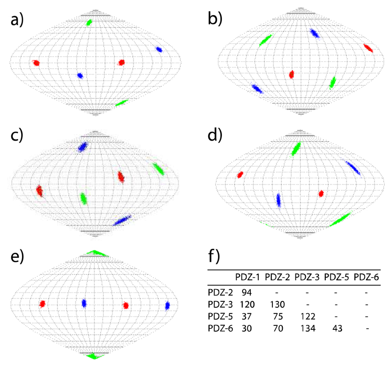
Figure 3.
Orientations of the alignment tensor transmitted to the 42 kDa protein MBPTGWETWV by the CLaNP-5 tagged PDZ mutants 1, 2, 3, 5, 6 (a-e). z, y and x axis are shown in red, blue and green in Sanson Flamsteed projections, respectively. The orientation of the alignment tensors was obtained by fitting the experimental RDCs to MBP’s 3D structure (PDB code: 1DMB).[21a] Uncertainties were evaluated by 1000 cycles of the "structural noise Monte-Carlo method".[16, 23] f) 5D angles between the alignment tensors induced in MBP.

