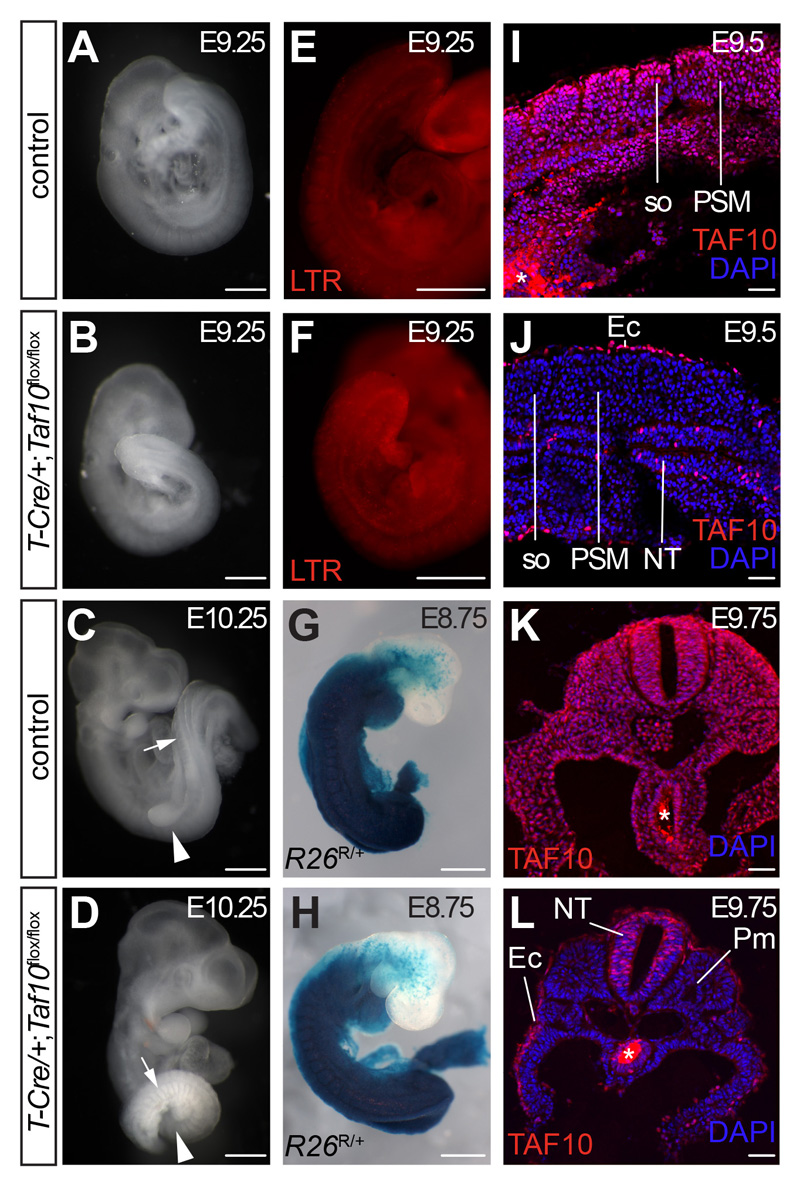
Fig. 4. Efficient Taf10 conditional deletion in the paraxial mesoderm.
(A-C) Whole-mount right-lateral view of control (A,C) and T-Cre;Taf10 mutant (B,D) E9.25 embryos (A,B) and E10.25 (C,D). The arrowhead in C,D indicates the position of the forelimb bud that is absent in the mutant, the arrow indicates the somites. (E,F) Cell death assay by LysoTracker Red (LTR) staining of E9.25 control (E) and T-Cre;Taf10 mutant (F) embryos. (G,H) Whole-mount X gal stained E8.75 T-Cre/+;R26R/+ control (G) and T-Cre/+;R26R/+;Taf10flox/flox mutant (H) embryos showing the efficient early recombination within the paraxial mesoderm. (I-L) DAPI counterstained TAF10 immunolocalization on E9.5 sagittal (I,J) and E9.75 transverse (K,L) sections from control (I,K) and T-Cre;Taf10 mutant (J,L) embryos. so; somites, PSM; presomitic mesoderm, NT; neural tube, Ec; ectoderm, Pm; paraxial mesoderm. The asterisk (*) in (K,L) indicates background due to secondary antibody trapping in the endoderm lumen. Scale bars: and I-L represent 500 µm in A-H and 50 µm in I-L.

