
Figure 1.
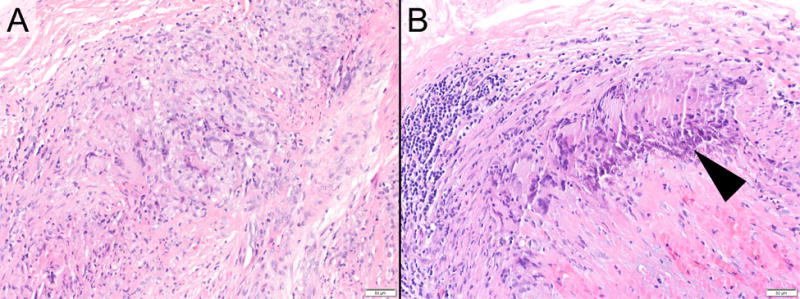
Example Photomicrographs exhibiting (A) a temporal artery with active granulomatous arteritis at the time of initial biopsy, and (B) ongoing active granulomatous arteritis at 6 months, post biopsy (hematoxylin and eosin staining; original magnifications, x200). Early calcification is also apparent in the follow-up sample (arrowhead).