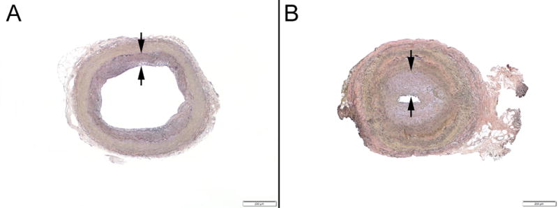
Figure 4.

Example photomicrographs showing (A) mild intimal fibroplasia (<25% luminal narrowing, between arrows) and (B) marked intimal fibroplasia (>75% luminal narrowing, between arrows) (Verhoeff-van Giesson staining; original magnifications, x40).
