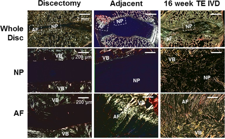
Fig 6. Picrosirius red-stained histology under polarized light showed birefringent features associated with collagen fibers.
Discectomy samples show collagen organization in vertebrae, with no tissue in the intervertebral space. Adjacent motion segments show the absence of collagen fibers in the NP and large collagen fibers (~50–100 μm) inserting from the AF to the vertebral body (VB). At 16 weeks, TE-IVD samples show the presence of some small, unorganized collagen fibers in the NP, with larger (~20–50 μm) fibers that insert into the vertebral body (VB).

