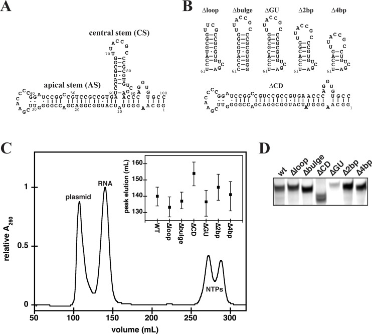
Fig 1.
(A) Secondary structure of adenovirus VAIΔTS (wt). (B) Schematic (not experimentally determined) representation of mutations in the CS of wt RNA and the ΔCD mutant that lacks the CS. (C) Purification of wt RNA by size exclusion chromatography (HiLoad 26/60 Superdex 75 column). Concentration of elution fractions was monitored by in-line spectrophotometric detection at 260 (solid line) and 280 nm simultaneously. The inset to the elution profile represents the elution range for the peak volume of each mutant RNA. (D) Native gel electrophoresis of wt RNA and its mutants. 2 μg of each RNA was loaded on 8% native TBE gel. Gels were stained with toluidine blue for total RNA.

