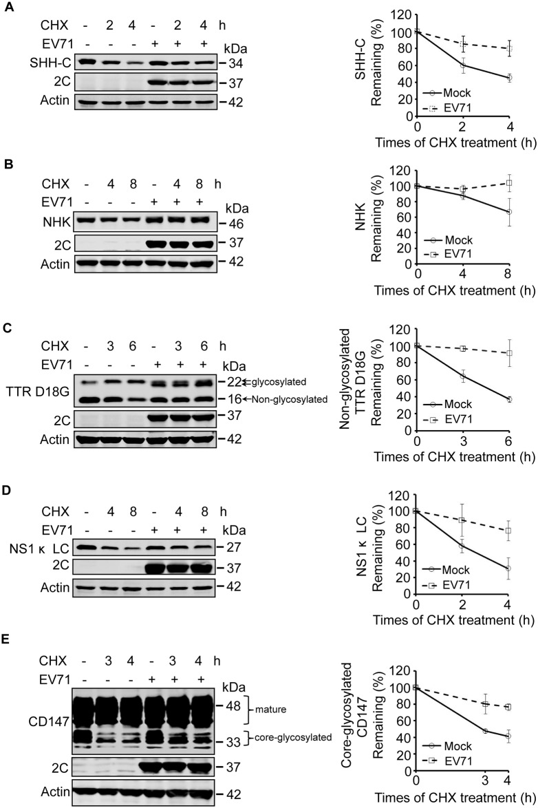
Fig 1. EV71 infection inhibits the degradation of ERAD substrates.
(A–D) RD cells stably expressing SHH-FLAG (A), NHK-FLAG (B), TTR D18G-FLAG (C), and NS1κ LC-FLAG (D) were mock-infected (−) or infected (+) with EV71 (MOI = 10) for different times. Nine hours post-infection the cells were treated with CHX (100 μg/ml) for the indicated times. The cell lysates were separated by SDS-PAGE and then analyzed by western blotting with indicated antibodies to detect the substrates, EV71 2C, and actin; actin was used as the loading control (left panel). The graph shows the quantification of the relative substrate (right panel). The data are presented as means ± SD of three independent experiments. (E) RD cells were treated as described in (A–D), and western blotting was used to detect the expression of CD147, EV71-2C, and actin (left panel). The graph shows the quantification of core-glycosylated CD147 (right panel). The data are presented as means ± SD of three independent experiments.

