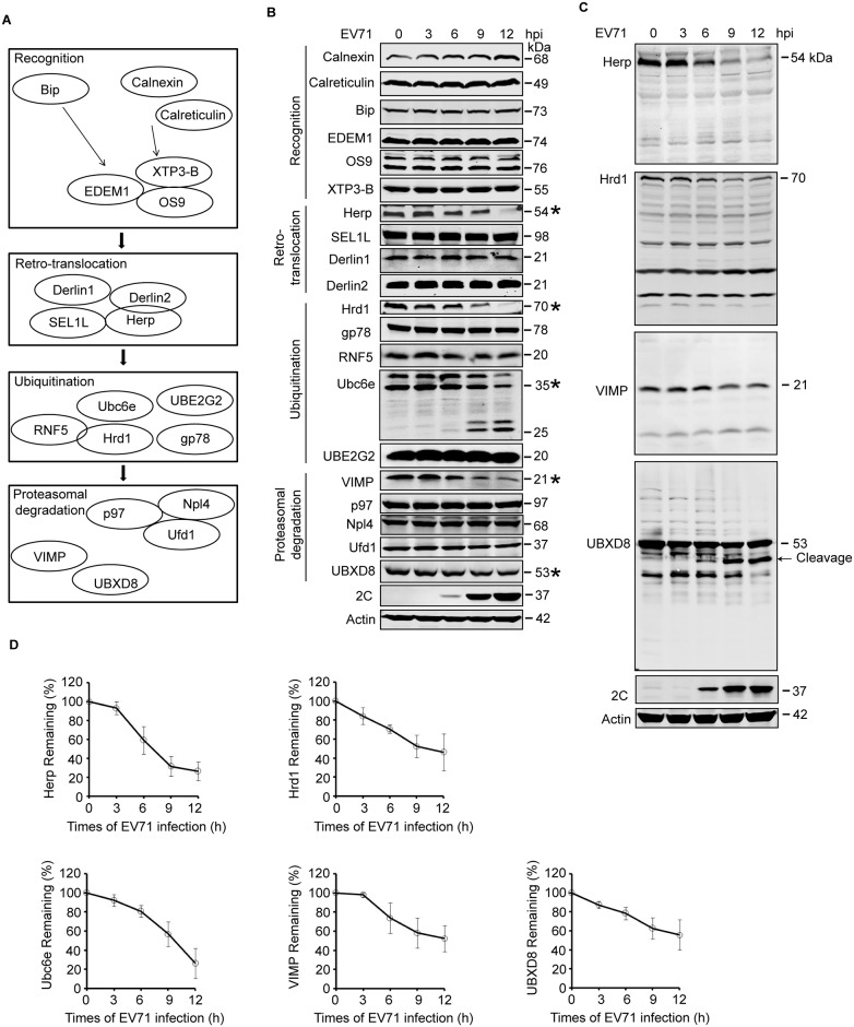
Fig 3. EV71 targets ERAD at multiple points.
(A) Diagram of the key molecules involved in ERAD. (B) RD cells were infected with EV71 (MOI = 10) for the indicated times (hpi: hours post-infection). The cells were then harvested and western blotting was performed using the indicated antibodies to detect the indicated ERAD components, EV71 2C, and actin. The ERAD molecules assessed in this study were separated into four categories: substrate recognition, retrotranslocation, ubiquitination, and proteasomal degradation. Asterisks indicate the molecules that were obviously downregulated. (C) Full-size western blots for Herp, Hrd1, VIMP, and UBXD8 described in (B). (D) Quantification of Ubc6e, Herp, Hrd1, VIMP, and UBXD8 in (B). The data are presented as means ± SD of three independent experiments.

