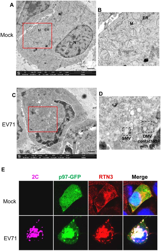
Fig 9. EV71 induces ROs in infected cells and redistributes RTN3 to viral ROs during infection.
(A) Low-magnification electron micrographs of mock infected RD cells. N, nucleus; M, mitochondria; ER, endoplasmic reticulum. Scale bar, 1 μm. (B) Magnified view of the boxed region in (A). (C) Low-magnification electron micrographs of EV71-infected RD cells (MOI = 10, 12 hpi). Scale bar, 1 μm. (D) Magnified view of the boxed region in (C). SMV, single membrane vesicle; DMV, double membrane vesicle. (E) RD cells stably expressing p97-GFP were mock-infected (upper panel) or infected with EV71 (MOI = 10) for 12 h (lower panel). The cells were then fixed and stained with rabbit anti-RTN3 and mouse anti-2C antibodies (p97, green; RTN3, red; 2C, purple; nuclei, blue).

