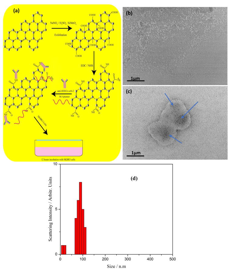
Figure 1.
(a) Schematics of synthesis and bioconjugation protocol of water soluble and biocompatible Graphene oxide from graphite powder. (b) Transmission electron microscope image of synthesized nanometer size two-dimensional graphene oxide soluble in aqueous media. (c) Transmission electron microscope image showing a significant binding and accumulation of S6 RNA aptamer and anti-HER2/c-erb-2 functionalized GO on the SKBR3 cell surface. (d) Size distribution of synthesized graphene oxide nanoparticle measured by the dynamic light scattering technique for monodisperse ~100 nm size.

