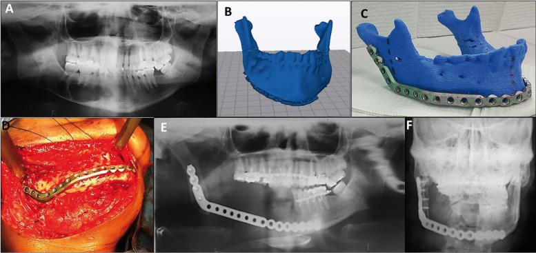
Figure 2.
(A). Panorex x-ray showing a multilocular lesion, approximately 5.0 x 2.5 cm in size in the right mandible (B, C). MRP model created from patient DICOM data and reconstruction plate (2.7-mm mandible reconstruction plate (KLS Martin®, Jacksonville, FL, USA) prebended on its corresponding RMP. (D). Submandibular flap exposing the right mandible and tumor. (E, F) Postoperative panoramic and skull x-ray showing correct positioning of the reconstruction plate and mandibular symmetry.

