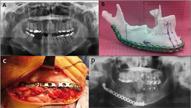
Figure 3.
(A) Panorex revealing a mixed multilocular lesion that was approximately 6 x 2 cm in size mainly in the right mandibular body. (B) MRP model created from patient DICOM and its corresponding prebended plate. (C) Submandibular flap with plate and bone graft in place. (D) Postoperative panorex showing correct positioning of the reconstruction plate.

