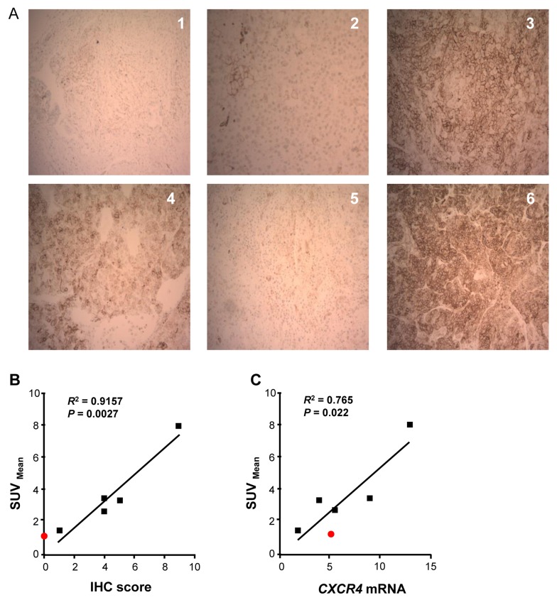
Figure 5. Uptake of 64Cu-plerixafor correlates with expression of CXCR4.
A. IHC staining of sections from the six resected pulmonary nodules. Nodule 1 was a chondroma and nodules 2-6 were metastatic ACC. Magnification is X 100. B. Linear regression analysis of CXCR4 IHC score vs. 64Cu-plerixafor SUVmean for the five excised ACC nodules. C. Linear regression analysis of CXCR4 mRNA vs. 64Cu-plerixafor SUVmean for the five excised ACC nodules. After normalization to measurements of 18S rRNA, values for CXCR4 mRNA were normalized to the value for the H295R cell line as in Figure 2. Red symbol in B and C corresponds to nodule 1 (chondroma), which was not included in statistical analyses. R2 is the coefficient of determination.

