Figure 7.
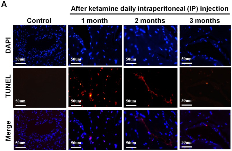
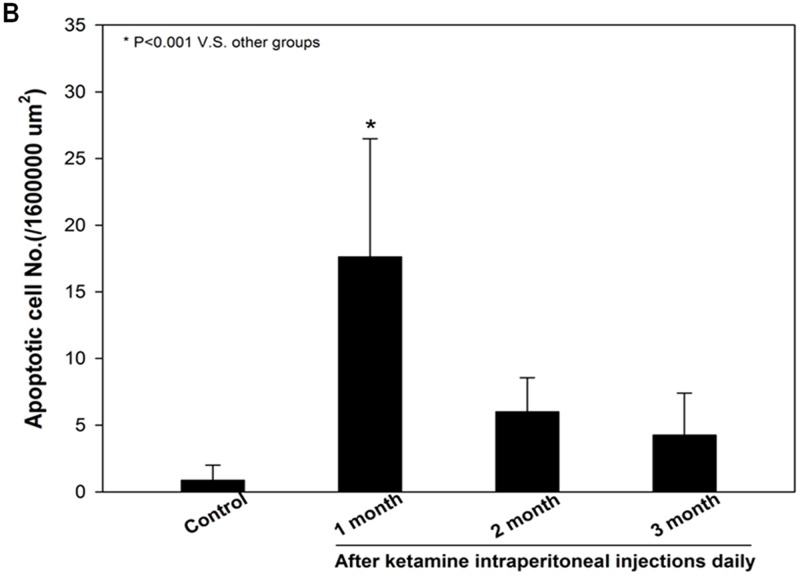
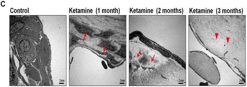
A. Transferase-mediated dUTP-biotin nick end labeling (TUNEL) staining from rats without or with ketamine treated for 1, 2 and 3 months showed nuclear colocalization with 4′, 6-diamidino-2-phenylindole (DAPI) (original magnification 200×). Only cells positive for both TUNEL and DAPI were considered positive for apoptosis. B. Ultrastructural analysis of the corpus cavernosum tissue from rats without or with ketamine treated for 1, 2 and 3 months. We observed abnormal structures and apoptosis of the smooth muscle cells at 1 month after ketamine treated and collagen deposition were clearly observed increase after ketamine treated 3 months. Arrow is corporal smooth muscle site; Arrow head is the collagen deposition (original magnification 5000×). C. Apoptotic cell quantification expressed as the number of apoptotic cell/area of the corpus cavernosum. Apoptotic index in ketamine treated 1 month significantly increase increased compared with that in others groups. *p < 0.001 compared with other groups.


