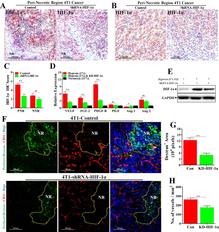
Figure 6. Knock-down of HIF-1α inhibited hypoxia-induced abnormal angiogenesis and reduced vessel leakage.
4T1 cancer cells transfected with Lentivirus-shRNA-HIF-1α or Lentivirus-shRNA-Con were orthotopically transplanted into the BALB/c mice. (A–C) IHC staining for HIF-1α, revealing a significant reduction of HIF-1α protein level in both (A) peri-necrotic and (B) non-necrotic regions of shRNA-HIF-1α 4T1 tumors; (C) quantification of HIF-1α IHC score (addition of intensity score and positive signal area). n = 9; scale bar: 100 μm. (D) Reduced relative mRNA expressions of various angiogenesis-associated factors in shRNA-HIF-1α 4T1 cancer cells cultured in the hypoxic condition (1% O2). n = 6. (E) Immunoblotting for HIF-1α, showing reduced HIF-1α protein level in Lentivirus-shRNA-HIF-1α 4T1 cancer cells cultured in hypoxic condition (1% O2) in vitro. 100 μg cellular protein was loaded on each lane. (F) Staining for fitc-conjugated dextran (green, 70 kD) and CD31 (red) in 4T1 tumors transfected with Lentivirus-shRNA-HIF-1α or Lentivirus-shRNA-Con. Before extraction, fitc-conjugated dextran was intravenously injected into the tumor-bearing mice for observation of vascular leakage. NR indicates necrotic tumor region. Scale bar: 100 μm. (G and H) Quantification of (G) detran+ area leaking outside the tumor lumen and (H) microvessel density (No. of vessels per mm2) in 4T1 tumors transfected with either Lentivirus-shRNA-HIF-1α or Lentivirus-shRNA-Con (n = 10). Quantitative data are indicated as mean ± SEM. *p < 0.05; **p < 0.01; ***p < 0.001; ns indicates no significant (P > 0.05).

