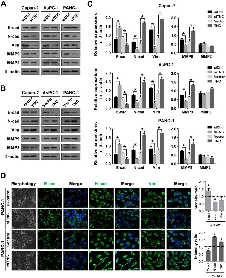
Figure 2. TNC regulates tumour metastasis-related genes in pancreatic cancer cells.
(A) Western blot analysis was performed to test the expression of epithelial (E-cadherin) and mesenchymal markers (N-cadherin, Vimentin) and MMPs (MMP9 and MMP2) in the Capan-2, AsPC-1 and PANC-1 cells transfected with siTNC or siControl. (B) The protein levels of EMT markers and MMPs were detected by western blot in Capan-2, AsPC-1 and PANC-1 cells after transfection with TNC plasmid or control vector. (C) The protein levels of E-cadherin, N-cadherin, Vimentin, MMP9 and MMP2 were normalized to β-actin in Capan-2, AsPC-1 and PANC-1 cells after the indicated treatment. (D) Cell morphology and confocal microscopy images of PANC-1 cells transfected with siTNC or coaded with rhTNC, as well as their controls. The green signal represents staining corresponding to E-cadherin, N-cadherin or Vimentin, and the blue signal represents the nuclear DNA staining by DAPI. Scale bar, 20 μm. The fluorescent intensity was determined and standardised to the cellular background. The levels of E-cad, N-cad and Vim were calculated as a ratio with compared to the controls. Data represent the mean ± SD.

