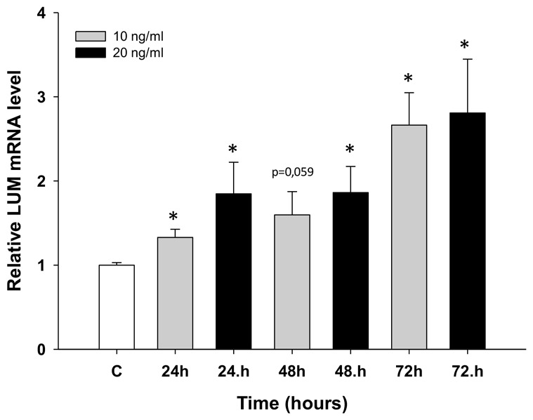
Figure 6. Expression analysis (Q-PCR) of the LUM gene in the W1 cell line after TOP treatment.

This figure presents relative gene expression in treated cells (TOP – 10 ng/ml: grey bars, TOP – 20 ng/ml: black bars) with respect to untreated cells (white bars) at different time points. Untreated cells were assigned a value of 1. Values were considered significant at *p < 0.05.
