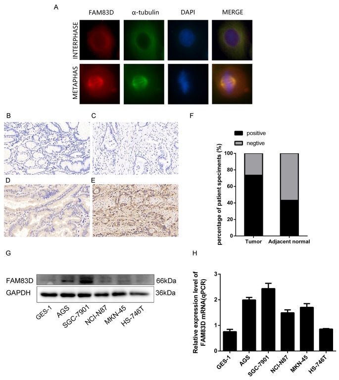
Figure 1. Expression of FAM83D in human gastric tumor tissues and cell lines.
(A) Immunofluorescence images of AGS cells at interphase and metaphase stained with antibodies against FAM83D (red), α-tubulin (green), and DAPI (blue). (B) Negative FAM83D expression in the non-tumor gastric mucosa (200x). (C, D, and E) Negative, weak positive and strong positive FAM83D expression in gastric cancer tissues. (F) Positive ratios of FAM83D expression in 102 pairs of gastric cancer tissues. (G and H) mRNA and protein expression of FAM83D in GC cell lines.

