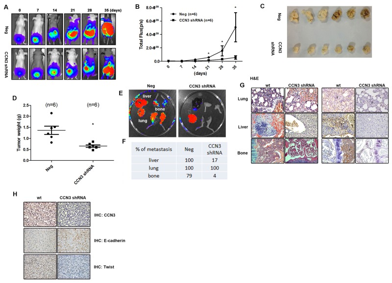
Figure 1. CCN3 is required for metastasis of PCa cells in mouse orthotopic model.
(A and B) CCN3 shRNA (CCN3 shRNA), and control vector PC3 cells (Neg) which stably expressed luciferase were used. The prostates of male nu/nu mice (6–8 weeks old) were exposed by midventral incision and injected with 5 × 10⁵ cells suspended in 50 μL culture media. One week after injection, surgical staples were removed, and the tumor growth and local metastasis were monitored using IVIS Imaging System. The panels depict quantification of fluorescence imaging data acquired at day 1, 7, 14, 21, 28, and 35. (C and D) The orthotopic model of nu/nu mice was used. The mice were sacrificed 28 days later, and the tumors were collected from injection site. The tumors were weighed and photographed. (E) The orthotopic model of nu/nu mice was set up. The mice were sacrificed 28 days later and their lungs, livers, and legs were dissected and monitored using IVIS Imaging System. (F) Quantification of fluorescence imaging data acquired by IVIS Imaging System in (E). (G) Left, hematoxylin and eosin (H&E) staining of lung, liver and limb from a control and CCN3 shRNA mice. Tumors were indicated as T. Right, the lung, liver, and limb specimens from sacrificed mice were stained with CCN3 antibody. (H) The tumor specimens from sacrificed mice were stained with CCN3, E-cadherin and N-cadherin antibodies. The stained specimens were photographed by optical microscope. Results are expressed as the mean ± S.E.M. *p < 0.05 compared with day 0.

