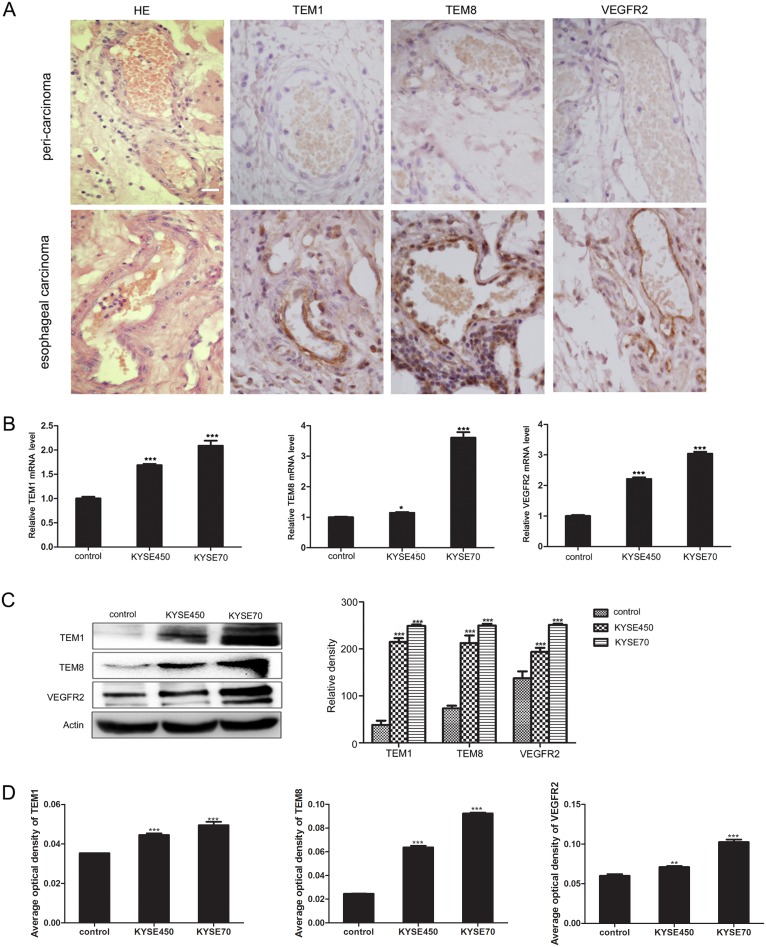
Figure 2. KYSE450 or KYSE70 TCM induced NECs to have the characteristics of TECs.
(A) Immunohistochemistry validated the TECs markers (TEM1, TEM8 and VEGFR2) in esophageal carcinoma and peri-carcinoma tissue. TEM1, TEM8 and VEGFR2 were preferentially expressed in vascular endothelial cells of esophageal carcinoma tissue (scale bar 20 μm). (B-D) NECs were induced by KYSE450 or KYSE70 TCM for 48 h. The relative mRNA levels of TECs markers were examined by qRT-PCR (B). The protein levels of TECs markers were detected by Western blot (C) and immunofluoresence (D). Results are expressed as mean ± SD. * p < 0.05, ** p < 0.01, *** p < 0.001.

