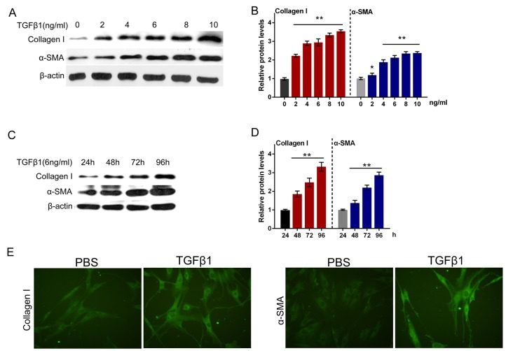
Figure 5. TGFβ1 effectively induced fibrosis process in primary human oral submucosa fibroblasts.
(A and B) The protein levels of Collagen I and α-SMA under a series of doses of TGFβ1 treatment (0, 2, 4, 6, 8, 10 ng/ml), as determined using Western blot assays. (C and D) The protein levels of Collagen I and α-SMA under 6 ng/ml TGFβ1 treatment for 24, 48, 72, 96 h, as determined using Western blot assays. The data are presented as mean ± SD of three independent experiments. **P<0.01, vs 0 ng/ml TGFβ1 group. (E) The immunofluorescence assays were performed to validate Collagen I and α-SMA expression of oral submucosa fibroblasts response to TGFβ1 (6 ng/ml for 48 h).

