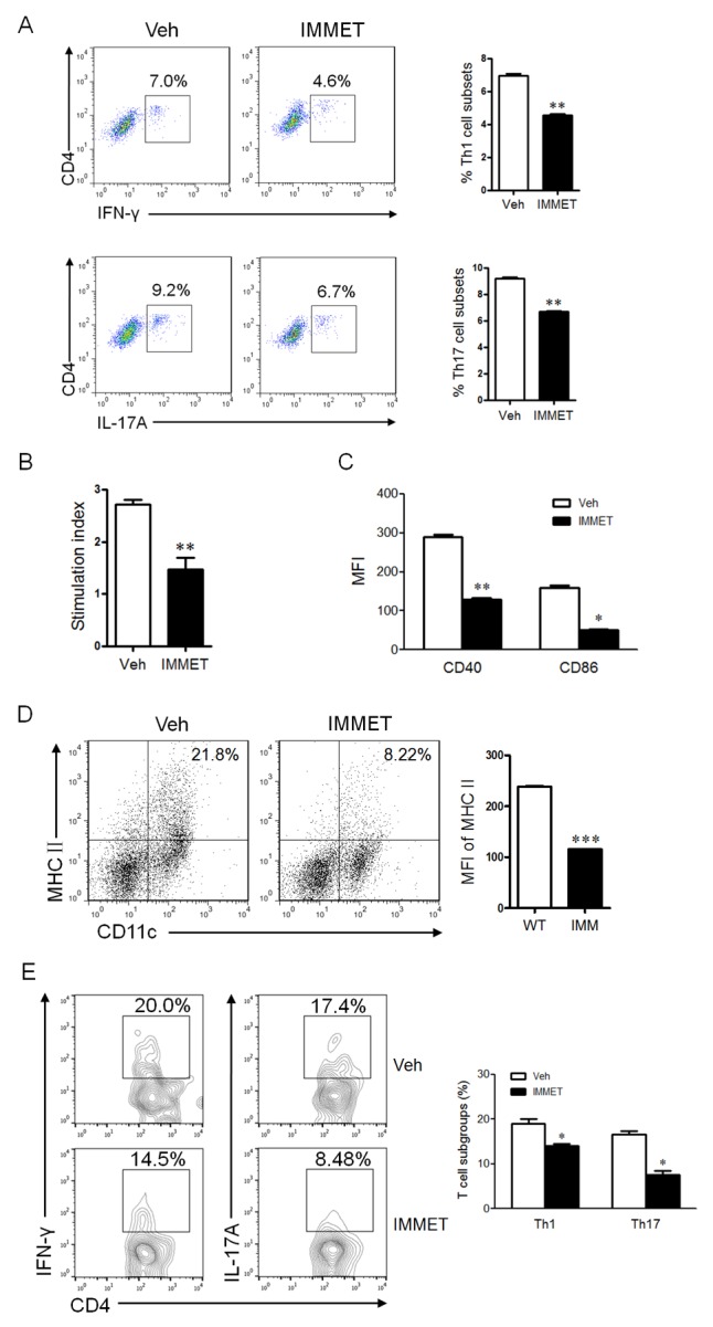
Figure 2. The treatment of immethridine could affect the differentiation and function of immune cells in EAE mice.

(A) FACS analysis of CD4+ T cell subsets in splenocytes of EAE mice (n=6). Splenocytes were prepared and Th1 and Th17 cells were analyzed by intracellular staining of IFN-γ and IL-17A, respectively, in the CD4+ gate. *p<0.05, **p<0.01 versus vehicle. (B) MTT analysis of MOG35-55-specific T cell proliferation. Splenocytes of EAE mice (n=6) were isolated and cultured at the present or absent of MOG35-55 peptides for 72h and T cell proliferation was measured using a MTT Cell Proliferation Assay Kit. The data were shown at stimulation index. **p<0.01 versus vehicle. (C) FACS analysis of expression of co-stimulatory molecules on DCs in splenocytes of EAE mice (n=6). Splenocytes were prepared and stained with fluorescent labeled-antibodies against CD11c, CD40 or CD86, the stained cells were analyzed on flow cytometry. *p<0.05, **p<0.01 versus vehicle. (D) FACS analysis of expression of MHCII on DCs in spinal cord of EAE mice (n=6). Immune cells in spinal cord were prepared and stained with fluorescent labeled-antibodies against CD11c, MHCII, the stained cells were analyzed on flow cytometry. ***p<0.001 versus vehicle. (E) FACS analysis of CD4+ T cell subsets in spinal cord of EAE mice (n=6). The lymphocytes from spinal cord were prepared and Th1 and Th17 cells were analyzed by intracellular staining of IFN-γ and IL-17A, respectively, in the CD4+ gate. *p<0.05 versus vehicle. Data are representative of three independent experiments.
