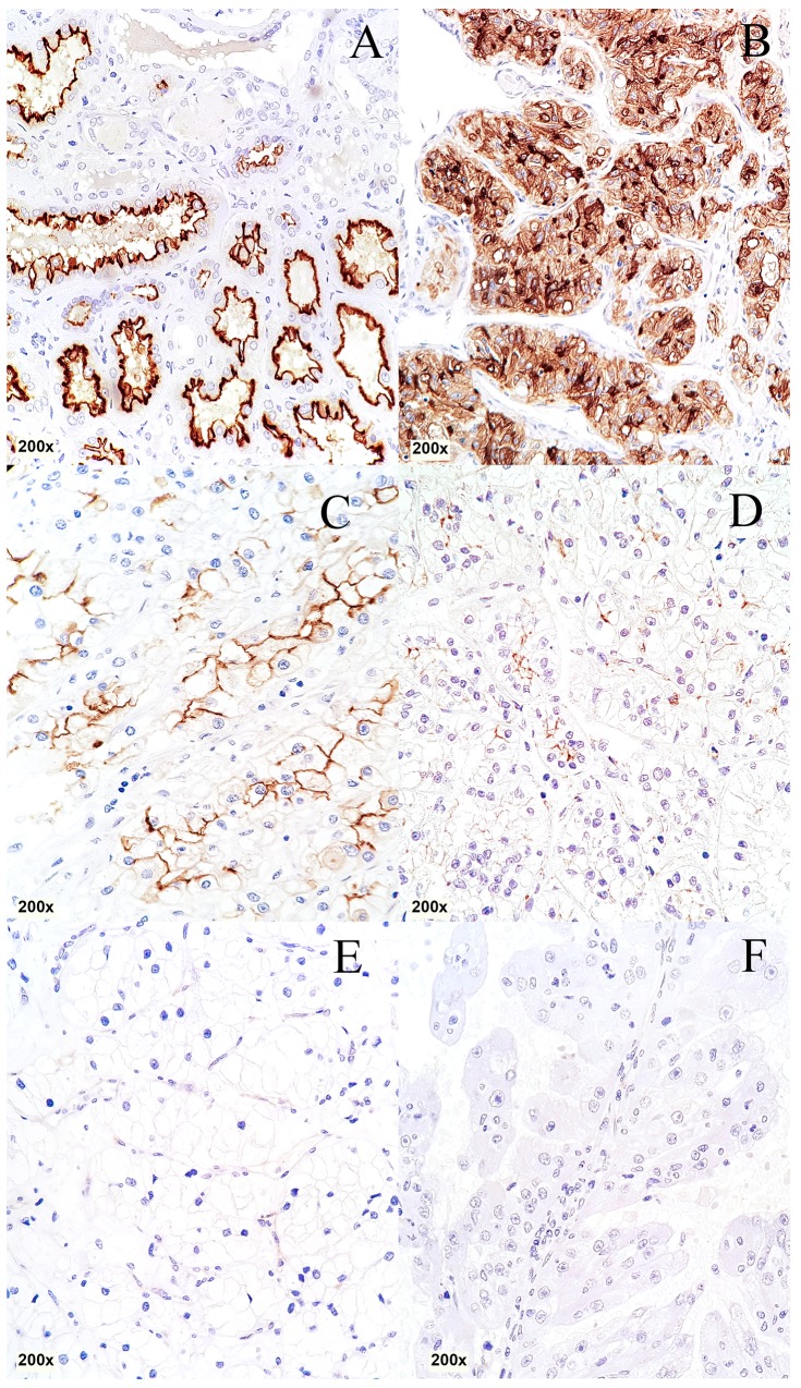
Figure 1. Immunohistochemical staining of CDHR5 in different histological subtypes of RCC.
(A) Normal kidney, (B) clear cell RCC: positive “3+”, (C) clear cell RCC: positive “2+”, (D) clear cell RCC: positive “1+”, (E) clear cell RCC: negative “0”, (F): papillary RCC: negative “0”. (total magnification for every image 200x)

