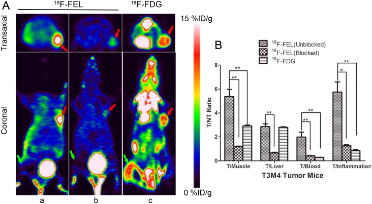
Figure 4.
(A) PET imaging of T3M4 tumor-bearing inflammation nude mice 60 min after injection of 18F-FEL (a) and 18F-FDG (c). PET images 60 min after co-injection of β-D-lactose (15 mg/kg) (b). (B) The uptake ratio of: tumor-to-organ were summarized. Note: * = P < 0.05, ** = P < 0.01. (Tumors are marked by red arrows and inflammatory lesions are marked by green arrows).

