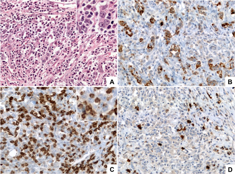
Figure 3. Lymphoepithelioma-like (LEL) carcinoma.
(A) (HE, 400×) shows a poorly differentiated carcinoma formed by medium to large size tumor cells with vesicular nuclei, prominent nucleoli and small amount of eosinophilic cytoplasm (inset) arranged in small clusters and poorly formed cords, intermingled with numerous lymphocytes. (B) (Pan-Cytokeratin AE1/AE3, 400×) shows that the tumor cells are positive for cytokeratins demonstrating their epithelial nature and highlighting the poorly formed clusters and cords arrangement. (C) (CD3, 400×) highlights the diffuse lymphocytic infiltrate composed mainly by mature T lymphocytes; tumor cells are negative for CD3 (inset). (D) (CD20, 400×) highlights scattered B-lymphocytes within the infiltrate.

