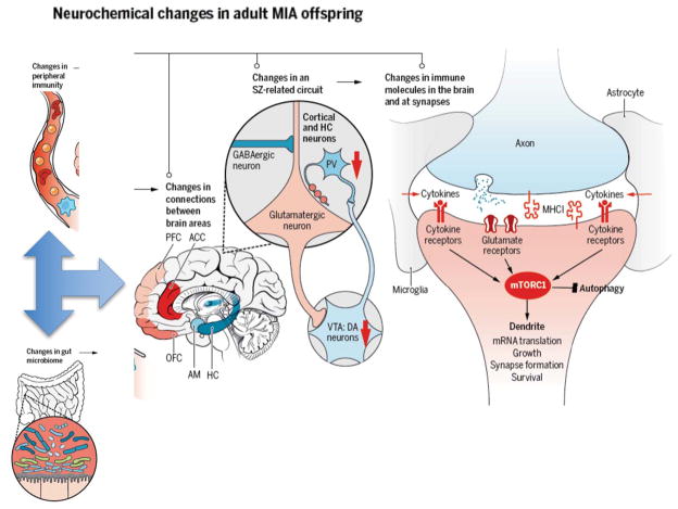
Figure 2. Mechanisms underlying the effects of MIA on brain function.
Aberrations in the microbiome following MIA can lead to altered development of peripheral immunity, which in turn leads to alterations in brain development. Deficits in long-range connections between brain regions implicated in SZ and ASD, including the hippocampus (HC), prefrontal cortex (PFC), the anterior cingulate cortex (ACC), the orbitofrontal cortex (OFC) and the amygdala (AM) have been reported in MIA(3, 4, 14). Specific alterations in activity of glutamatergic neurons in the cortex caused by decreased function of dopaminergic neurons in the ventral tegmental area (VTA) and decreased GABAergic input also occurs in SZ and ASD as well as the MIA models(3, 4, 14). Changes in expression of immune molecules in the brain and even at synapses, including cytokines and MHCI molecules, alters synaptic plasticity and function, and contributes to the changes in circuitry and connectivity between brain regions that characterize these disorders(6). Finally, alterations in immune and neuronal signaling due to MIA may converge upon the mammalian target of rapamycin (mTOR) pathway, which regulates synapse formation, growth, translation, survival, and autophagy(68).

