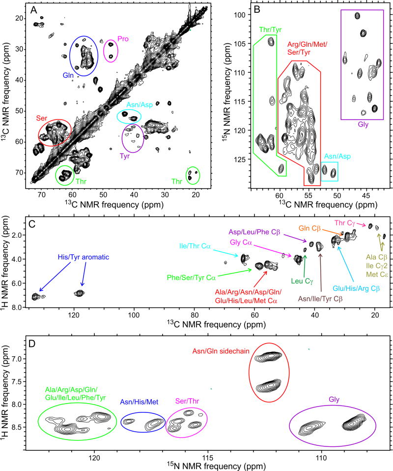
Fig. 2. Coexistence of structural order and dynamic disorder within FUS-LC fibrils.
(A,B) 2D 13C-13C and 15N-13C NMR spectra of U-FUS-LC fibrils, recorded under conditions that select for signals from immobilized, structurally ordered segments. (C,D) 2D 1H- 13C and 1H-15N NMR spectra of U-FUS-LC fibrils, recorded under conditions that select for signals from highly flexible, dynamically disordered segments. Residue-type assignments are based on characteristic chemical shift ranges. Contour levels increase by successive factors of 1.40, 1.35, 1.80, and 1.40 in (A), (B), (C), and (D), respectively. See also Figs. S2 and S3 and Table S3.

