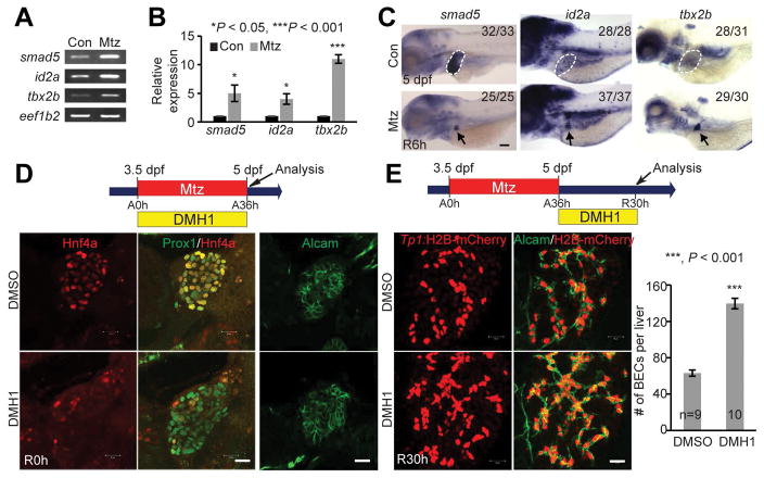
Figure 1. Bmp signaling is required for BEC-driven liver regeneration.
(A, B) RT-PCR (A) and qPCR (B) data showing the expression levels of smad5, id2a and tbx2b between control and regenerating livers at R6h. (C) WISH images showing the expression of smad5, id2a and tbx2b in control (dashed lines) and regenerating (arrows) livers. Numbers indicate the proportion of larvae exhibiting the expression shown. (D) Single-optical section images showing the expression of Hnf4a and Prox1 or Alcam in regenerating livers. (E) Confocal projection images showing Tp1:H2B-mCherry and Alcam expression in regenerating livers. Quantification of the number of H2B-mCherry/Alcam double-positive cells (i.e., BECs). Scale bars: 150 (C), 20 (D,E) μm; error bars: ±SEM.

