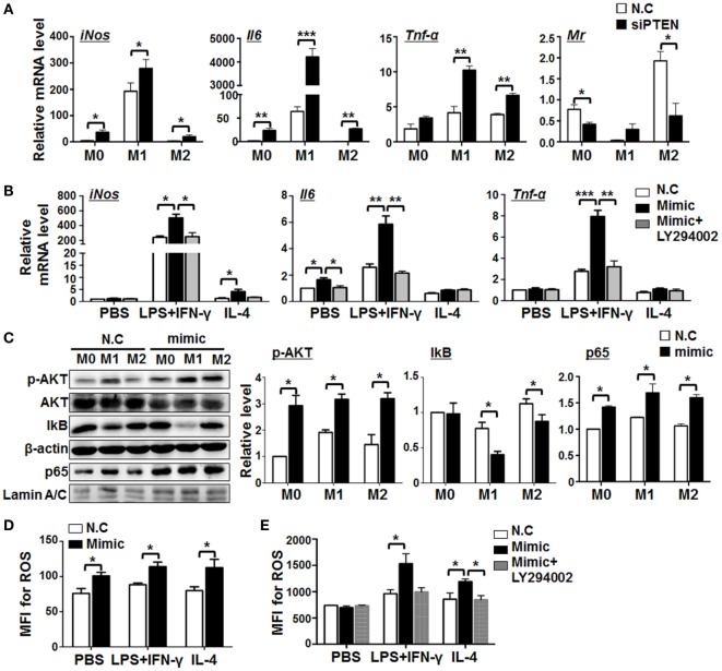
Figure 6.
miR-148a-3p promotes M1 macrophage polarization via PTEN/AKT/NF-κB signaling. (A) BM-derived macrophages (BMDMs) were transfected with siRNA targeting Pten (siPTEN) or N.C., and then polarized with PBS (M0), lipopolysaccharide (LPS) + interferon (IFN)-γ (M1), or interleukin (IL)-4 (M2). mRNA levels of iNos, Il6, Tnf-α, and Mr were determined by qRT-PCR, using β-actin as a reference control (n = 3). (B) BMDMs were transfected with miR-148a-3p mimic or N.C., and then polarized with PBS (M0), LPS + IFN-γ (M1), or IL-4 (M2), in the presence of the AKT inhibitor, LY294002. The mRNA levels of iNos, Il6, and Tnf-α were determined by qRT-PCR, using β-actin as a reference control (n = 3). (C) BMDMs were transfected with miR-148a-3p mimic or N.C., and then polarized with PBS (M0), LPS + IFN-γ (M1), or IL-4 (M2). The levels of phosphorylated AKT (p-AKT), total AKT, IκB, and nuclear p65 proteins were determined by Western blotting, using β-actin or Lamin A/C as reference controls, and quantitatively compared (n = 3). (D) BMDMs were transfected with miR-148a-3p mimic or N.C., and then polarized with PBS (M0), LPS + IFN-γ (M1), or IL-4 (M2). Reactive oxygen species (ROS) levels were determined using FACS (Figure S4A in Supplementary Material) and expressed as mean fluorescent intensity (MFI) (n = 3). (E) BMDMs were transfected with miR-148a-3p mimic or N.C., and then polarized with PBS (M0), LPS + IFN-γ (M1), or IL-4 (M2), in the presence of LY294002. ROS levels were determined using FACS (Figure S4B in Supplementary Material) and are expressed as MFI (n = 3). Bars, mean ± SD; *P < 0.05; **P < 0.01; ***P < 0.001.

